Novai, a medtech company combatting blindness and disability by pioneering early detection and intervention for eye and brain diseases, has announced the publication of a peer-reviewed study in Expert Review of Ophthalmology, which successfully demonstrates its ‘Detection of Apoptosing Retinal Cells’ (DARC) imaging technology as a promising biomarker for identifying patients at high risk of central vision loss caused by macular atrophy (MA).
By revealing disease activity at an earlier stage than currently possible, DARC offers a novel approach to predicting future patterns of atrophy progression, supporting more accurate patient stratification for clinical trials, and earlier initiation of treatment, as appropriate.
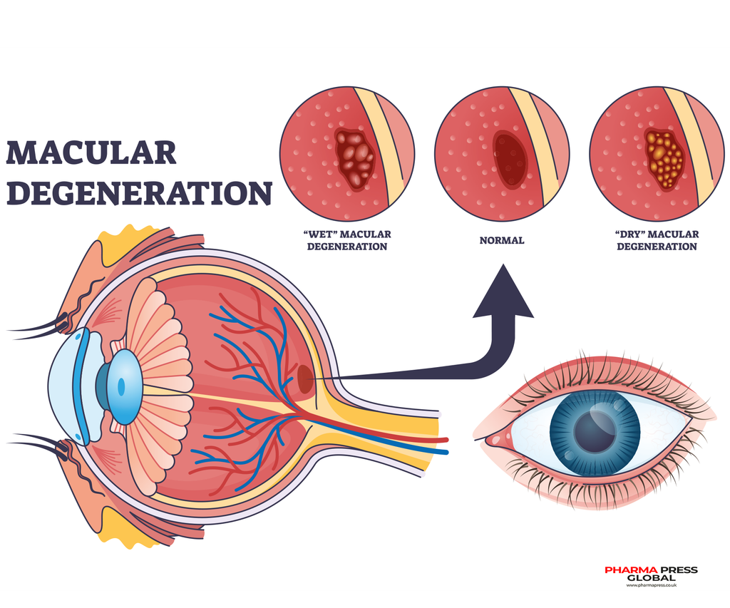
MA, also known as late dry-AMD or Geographic Atrophy (GA), is the advanced form of age-related macular degeneration (AMD) which is a leading cause of irreversible vision loss and blindness worldwide.
Characterised by the progressive loss of retinal cells responsible for central vision, a key challenge in MA is that retinal cells can become stressed and begin to die long before structural damage is visible on conventional imaging.
This has historically limited the ability to identify patients at greatest risk of vision loss. Novai’s DARC imaging technology combines a fluorescently labelled Annexin A5 probe with advanced data science, AI-powered algorithms, and digital image processing, to detect and quantify the number and pattern of distribution of sick, stressed, and apoptotic retinal cells in patients, before structural loss becomes apparent.
The paper, titled ‘Predicting directional macular atrophy progression using angular plot analysis of DARC (apoptosing retinal cells), EZ (ellipsoid zone) loss, EZ–RPE (retinal pigment epithelium) difference, and HRF (hyperreflective foci)’, builds on research carried out at Imperial College London, demonstrating the application of Novai’s DARC retinal imaging technology for the detection of early cellular stress, and predicting the directional expansion of atrophy.